
ABBA
ABBA+BraiAn: enhanced whole-brain histological mapping
In neuroscience, microscopy is essential for understanding the architecture and function of the brain. Mostly performed primarily in animal models, although also used on postmortem human tissue, this approach involves sectioning the brain into thin slices and imaging them at high resolution. With the recent advent of novel imaging technologies, it is now possible to scale this approach from individual sections to the entire brain. Although this represents a major breakthrough, it also introduces new challenges for image analysis that require automation.
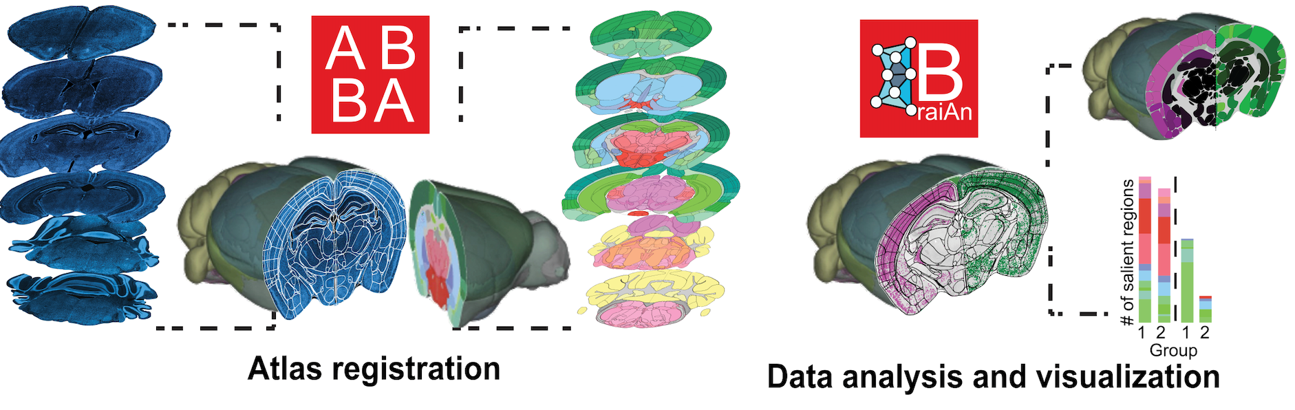
First, we need automated methods to locate and identify brain regions across large-scale microscopy dataset (brain atlas registration challenge). Second, we need to automate the quantitative analysis of signals of interest.
As an experimental neuroscience laboratory, we encountered these challenges firsthand and identified a clear gap in the available solutions. By surveying the field, we realized that this need is shared by thousands of researchers worldwide.
To address this gap, we collaborated with EPFL to develop ABBA+BraiAn, a unified software suite designed to support every step of whole-brain histological analysis, from 2D-to-3D atlas registration to signal quantification, data analysis, and visualization.
In its current version, ABBA+BraiAn provides a robust, versatile, and user-friendly solution for whole-brain analysis and has been widely adopted by the neuroscience community. With the support of the MSI, we will be able to further improve the robustness and accessibility of ABBA+BraiAn, making it more useful and accessible to researchers worldwide.
Project Leader
Project Participants
- Marco Milanesio, MSI.
- Carlo Castoldi, IPMC, Inria.